Modelling the eye: The cornea
We start at the very front of the eye. What shape is the cornea, why isn't it a simple sphere, and how do we build it in WebGL?
Ask a graphics programmer to model an eyeball and the first instinct is usually a glass sphere. It renders fast and looks clean. The problem is that a real eye shaped like a perfect sphere would turn every night-time streetlight into a glowing smear.
The clear dome at the front of the eye, the cornea, is a more interesting shape than a sphere, and it does most of the focusing. Of the eye’s roughly 60 diopters of optical power, the cornea supplies about 43. The famous crystalline lens inside handles the rest and does the fine-tuning. But how do we know it is not just a solid dome of biological glass? When Antonie van Leeuwenhoek first looked at corneal tissue through his homemade microscopes in the 1680s, he found it was not glass at all, but a tightly woven fabric of living fibers.
In this series we build the eye one part at a time in WebGL. Each section below has a live 3D view, and a Code tab with the full source. Let us start at the front.
The spherical cap: a first approximation
Before hospital scanners existed, people measured the eye with clever tricks. In 1619 Christoph Scheiner held glass marbles of known size next to a person’s eye and matched the reflections until the curvatures agreed. Two centuries of refinement later, we have the number every optometrist still uses: the front of the cornea has a radius of curvature of about R≈7.8 mm.
The visible cornea is a slightly oval window, roughly 11.7 mm wide and 10.6 mm tall, because the white of the eye overlaps it top and bottom. For a first WebGL model we ignore that and slice a clean cap off a sphere.
In Three.js a sphere already knows how to draw itself. We just tell it to keep only the front cap by limiting the vertical sweep angle:
// A cornea, roughly, is a cap sliced off the front of a sphere.
// The last two arguments keep only the top slice instead of the whole ball.
const geometry = new THREE.SphereGeometry(
  1,               // radius
  64, 64,          // horizontal and vertical smoothness
  0, Math.PI * 2,  // sweep the full circle around
  0, Math.PI / 4   // but only 45 degrees down from the top: the cap
);import React, { useEffect, useRef } from "react";
import * as THREE from "three";
import { OrbitControls } from "three/examples/jsm/controls/OrbitControls.js";
import { observeThreeResize } from "../threeResize";
export default function CorneaStep1() {
const mountRef = useRef<HTMLDivElement>(null);
useEffect(() => {
if (!mountRef.current) return;
// Scene setup
const scene = new THREE.Scene();
// Camera setup
const camera = new THREE.PerspectiveCamera(45, 1, 0.1, 100);
camera.position.set(-0.176, -0.038, 2.126);
camera.zoom = 1;
// Renderer setup
const renderer = new THREE.WebGLRenderer({ antialias: true, alpha: true });
renderer.setPixelRatio(window.devicePixelRatio);
renderer.setClearColor(0x000000, 0); // Transparent background
mountRef.current.appendChild(renderer.domElement);
// Controls setup
const controls = new OrbitControls(camera, renderer.domElement);
controls.enableDamping = true;
controls.dampingFactor = 0.05;
controls.target.set(-0.734, 0.003, -0.081);
const resizeObserver = observeThreeResize(
mountRef.current,
renderer,
camera,
);
// Lighting
const ambientLight = new THREE.AmbientLight(0xffffff, 0.6);
scene.add(ambientLight);
const dirLight = new THREE.DirectionalLight(0xffffff, 1.5);
dirLight.position.set(5, 5, 5);
scene.add(dirLight);
// Geometry: Simple Sphere (Javal's early approximation)
// Radius of curvature ~7.8mm. Let's scale it down for our scene.
const radius = 1;
const widthSegments = 64;
const heightSegments = 64;
// We only want a cap. Phi starts at 0 (top), goes down.
// A cornea is roughly the anterior 1/6th of the eye.
const phiStart = 0;
const phiLength = Math.PI / 4;
const geometry = new THREE.SphereGeometry(
radius,
widthSegments,
heightSegments,
0,
Math.PI * 2,
phiStart,
phiLength,
);
// Material matching WebGLEye "vibe"
const material = new THREE.MeshStandardMaterial({
color: 0x52525b,
roughness: 0.9,
transparent: true,
opacity: 0.6,
depthWrite: false,
side: THREE.DoubleSide,
});
const mesh = new THREE.Mesh(geometry, material);
// Add wireframe layer for the high-quality tech vibe
const wireMaterial = new THREE.MeshBasicMaterial({
color: 0xa1a1aa,
wireframe: true,
transparent: true,
opacity: 0.4,
});
const wireMesh = new THREE.Mesh(geometry, wireMaterial);
mesh.add(wireMesh);
// Rotate so the apex (+Y of sphere) points left (-X)
mesh.rotation.z = Math.PI / 2;
scene.add(mesh);
// Animation Loop
let animationFrameId: number;
const animate = () => {
animationFrameId = requestAnimationFrame(animate);
controls.update();
renderer.render(scene, camera);
const debugEl = mountRef.current
?.closest(".interactive-viewer")
?.querySelector(".debug-output");
if (debugEl) {
const state = {
camera: {
position: {
x: Number(camera.position.x.toFixed(3)),
y: Number(camera.position.y.toFixed(3)),
z: Number(camera.position.z.toFixed(3)),
},
rotation: {
x: Number(camera.rotation.x.toFixed(3)),
y: Number(camera.rotation.y.toFixed(3)),
z: Number(camera.rotation.z.toFixed(3)),
},
zoom: Number(camera.zoom.toFixed(3)),
},
target: {
x: Number(controls.target.x.toFixed(3)),
y: Number(controls.target.y.toFixed(3)),
z: Number(controls.target.z.toFixed(3)),
},
};
(debugEl as HTMLElement).innerText = JSON.stringify(state, null, 2);
}
};
animate();
return () => {
cancelAnimationFrame(animationFrameId);
resizeObserver.disconnect();
if (mountRef.current && renderer.domElement.parentNode) {
mountRef.current.removeChild(renderer.domElement);
}
renderer.dispose();
geometry.dispose();
material.dispose();
};
}, []);
return (
<div className="relative w-full h-full">
<div
ref={mountRef}
className="w-full h-full min-h-[400px]"
style={{ cursor: "grab" }}
/>
</div>
);
}So why is a sphere a bad lens? Light rays hitting the steep edge of a sphere bend more sharply than rays near the center, so they focus at slightly different points. The result is spherical aberration: a bright point of light spreads into a soft halo instead of a crisp dot. To see well at night, the eye had to abandon the sphere.
The aspheric cornea: flattening the edges (the Q value)
Nature fixes spherical aberration by gently flattening the cornea toward its edges while keeping the center steep. Optical engineers describe this shape mathematically using conic sections (like ellipses or parabolas), governed by a single number: the conic constant Q. The height of the surface (called the sag, z) at a distance r from the center is defined as:
z(r)=1+1−(1+Q)c2r2cr2
Here c=1/R is the curvature at the very center. The value of Q changes the shape:
- Q=0 (sphere): constant curvature everywhere. Strong halos around night lights.
- Q<0 (prolate): steeper center, flatter edges. The healthy human cornea sits near Q≈−0.26.
- Q>0 (oblate): flatter center, steeper edges. Common after laser surgery, and it makes night glare worse.
Interestingly, the human cornea does not flatten all the way to the value that would cancel spherical aberration completely (about −0.53). It leaves a little behind on purpose, because the lens deeper in the eye bends light the opposite way and cancels most of what is left. The two roughly balance out in a young, healthy eye.
Translating the sag formula into code is almost a direct copy:
const c = 1 / R;                 // curvature at the apex
for (let r = 0; r <= maxR; r += step) {
  const root = 1 - (1 + Q) * c * c * r * r;
  const z = (c * r * r) / (1 + Math.sqrt(root)); // surface height at radius r
  // ...place a ring of vertices at this height
}
Use the buttons below to switch between the healthy human cornea, a perfect sphere, and a post-surgery oblate shape. The color runs from green at the center to the shape’s color at the rim, so you can see where each one bends light differently.
import React, { useEffect, useRef, useState } from "react";
import * as THREE from "three";
import { OrbitControls } from "three/examples/jsm/controls/OrbitControls.js";
import { observeThreeResize } from "../threeResize";

const R = 7.8;
const MAX_R = 5.2; // wide aperture so the edge flattening is easy to see

// Three shapes worth comparing, each a single conic constant Q.
const PRESETS = [
	{
		id: "human",
		label: "Normal human",
		q: -0.26,
		hex: 0x22d3ee,
		note: "Prolate: steep center, flatter edges. Cancels most spherical aberration.",
	},
	{
		id: "sphere",
		label: "Perfect sphere",
		q: 0.0,
		hex: 0xa1a1aa,
		note: "Constant curvature. Edge rays overbend, so night lights bloom into halos.",
	},
	{
		id: "lasik",
		label: "Post-LASIK",
		q: 0.6,
		hex: 0xf43f5e,
		note: "Oblate: flatter center, steeper edges. Amplifies glare after surgery.",
	},
];

function sag(r: number, q: number) {
	const c = 1 / R;
	const root = 1 - (1 + q) * c * c * r * r;
	return root >= 0 ? (c * r * r) / (1 + Math.sqrt(root)) : NaN;
}

function buildGeometry(q: number, hex: number) {
	const radialSegments = 72;
	const angularSegments = 72;
	const vertices: number[] = [];
	const colors: number[] = [];
	const indices: number[] = [];
	const base = new THREE.Color(hex);
	const apex = new THREE.Color(0x34d399);

	for (let i = 0; i <= radialSegments; i++) {
		const r = (i / radialSegments) * MAX_R;
		let z = sag(r, q);
		if (Number.isNaN(z)) z = vertices[vertices.length - 1] ?? 0;
		const col = apex.clone().lerp(base, r / MAX_R);
		for (let j = 0; j <= angularSegments; j++) {
			const theta = (j / angularSegments) * Math.PI * 2;
			vertices.push(r * Math.cos(theta), r * Math.sin(theta), z);
			colors.push(col.r, col.g, col.b);
		}
	}

	for (let i = 0; i < radialSegments; i++) {
		for (let j = 0; j < angularSegments; j++) {
			const a = i * (angularSegments + 1) + j;
			const b = a + 1;
			const c2 = (i + 1) * (angularSegments + 1) + j;
			const d = c2 + 1;
			indices.push(a, b, d);
			indices.push(a, d, c2);
		}
	}

	const geometry = new THREE.BufferGeometry();
	geometry.setAttribute(
		"position",
		new THREE.Float32BufferAttribute(vertices, 3),
	);
	geometry.setAttribute("color", new THREE.Float32BufferAttribute(colors, 3));
	geometry.setIndex(indices);
	geometry.computeVertexNormals();
	return geometry;
}

// The Q = 0 sphere profile in a plane, used as a fixed reference the shape
// visibly pulls away from at the edges.
function sphereProfile(planeYZ: boolean) {
	const pts: THREE.Vector3[] = [];
	for (let t = -MAX_R; t <= MAX_R; t += 0.1) {
		const z = sag(Math.abs(t), 0);
		pts.push(planeYZ ? new THREE.Vector3(0, t, z) : new THREE.Vector3(t, 0, z));
	}
	return new THREE.BufferGeometry().setFromPoints(pts);
}

export default function CorneaAspheric() {
	const mountRef = useRef<HTMLDivElement>(null);
	const [preset, setPreset] = useState(PRESETS[0]);
	const updateRef = useRef<((q: number, hex: number) => void) | null>(null);

	useEffect(() => {
		if (!mountRef.current) return;

		const scene = new THREE.Scene();
		const camera = new THREE.PerspectiveCamera(45, 1, 0.1, 100);
		camera.position.set(6, 4, 18); // three-quarter view that shows the edge profile

		scene.add(new THREE.AmbientLight(0xffffff, 0.7));
		const dirLight = new THREE.DirectionalLight(0xffffff, 0.9);
		dirLight.position.set(5, 10, 10);
		scene.add(dirLight);

		const renderer = new THREE.WebGLRenderer({ antialias: true, alpha: true });
		renderer.setPixelRatio(window.devicePixelRatio);
		renderer.setClearColor(0x000000, 0);
		mountRef.current.appendChild(renderer.domElement);

		const controls = new OrbitControls(camera, renderer.domElement);
		controls.enableDamping = true;
		controls.dampingFactor = 0.05;
		controls.target.set(0, 0, 0);

		const resizeObserver = observeThreeResize(
			mountRef.current,
			renderer,
			camera,
		);

		const material = new THREE.MeshStandardMaterial({
			vertexColors: true,
			roughness: 0.25,
			metalness: 0.1,
			transparent: true,
			opacity: 0.9,
			side: THREE.DoubleSide,
		});
		const wireMaterial = new THREE.MeshBasicMaterial({
			color: 0xffffff,
			wireframe: true,
			transparent: true,
			opacity: 0.12,
		});

		let geo = buildGeometry(preset.q, preset.hex);
		const mesh = new THREE.Mesh(geo, material);
		const wire = new THREE.Mesh(geo, wireMaterial);
		mesh.add(wire);
		scene.add(mesh);

		// Fixed dashed reference: where a perfect sphere (Q = 0) would sit.
		const refMat = new THREE.LineDashedMaterial({
			color: 0xffffff,
			transparent: true,
			opacity: 0.5,
			dashSize: 0.25,
			gapSize: 0.15,
		});
		[sphereProfile(false), sphereProfile(true)].forEach((g) => {
			const line = new THREE.Line(g, refMat);
			line.computeLineDistances();
			scene.add(line);
		});

		updateRef.current = (q: number, hex: number) => {
			const newGeo = buildGeometry(q, hex);
			mesh.geometry.dispose();
			mesh.geometry = newGeo;
			wire.geometry.dispose();
			wire.geometry = new THREE.WireframeGeometry(newGeo);
			geo = newGeo;
		};

		let animationFrameId: number;
		const animate = () => {
			animationFrameId = requestAnimationFrame(animate);
			controls.update();
			renderer.render(scene, camera);

			const debugEl = mountRef.current
				?.closest(".interactive-viewer")
				?.querySelector(".debug-output");
			if (debugEl) {
				const state = {
					camera: {
						position: {
							x: +camera.position.x.toFixed(3),
							y: +camera.position.y.toFixed(3),
							z: +camera.position.z.toFixed(3),
						},
					},
					target: {
						x: +controls.target.x.toFixed(3),
						y: +controls.target.y.toFixed(3),
						z: +controls.target.z.toFixed(3),
					},
				};
				(debugEl as HTMLElement).innerText = JSON.stringify(state, null, 2);
			}
		};
		animate();

		return () => {
			cancelAnimationFrame(animationFrameId);
			resizeObserver.disconnect();
			if (mountRef.current && renderer.domElement.parentNode) {
				mountRef.current.removeChild(renderer.domElement);
			}
			renderer.dispose();
			geo.dispose();
			material.dispose();
			wireMaterial.dispose();
			refMat.dispose();
			updateRef.current = null;
		};
		// eslint-disable-next-line react-hooks/exhaustive-deps
	}, []);

	useEffect(() => {
		updateRef.current?.(preset.q, preset.hex);
	}, [preset]);

	return (
		<div className="relative w-full h-[450px] md:h-[550px] bg-transparent overflow-hidden rounded-lg group">
			<div className="absolute top-4 left-4 z-10 flex flex-col items-start gap-1 pointer-events-none bg-card/80 backdrop-blur-md border border-border p-3 rounded-xl shadow-lg max-w-[240px]">
				<span className="text-[10px] uppercase tracking-wider font-bold text-muted-foreground border-b border-border/50 pb-1 w-full text-center mb-1">
					Asphericity (Q)
				</span>
				<div className="flex justify-between w-full text-xs">
					<span className="font-bold">Conic constant Q</span>
					<span
						className="font-mono font-bold"
						style={{ color: `#${preset.hex.toString(16)}` }}
					>
						{preset.q.toFixed(2)}
					</span>
				</div>
				<div className="text-[10px] text-muted-foreground mt-1">
					{preset.note}
				</div>
				<div className="flex items-center gap-2 text-[10px] text-muted-foreground mt-1 pt-1 border-t border-border/40 w-full">
					<span className="w-4 border-t border-dashed border-white/70 inline-block"></span>{" "}
					perfect sphere reference
				</div>
			</div>

			<div className="absolute bottom-4 left-1/2 -translate-x-1/2 z-20 flex gap-2 bg-card/80 backdrop-blur-md border border-border p-2 rounded-2xl shadow-xl">
				{PRESETS.map((p) => (
					<button
						key={p.id}
						onClick={() => setPreset(p)}
						className={`px-3 py-1.5 rounded-lg text-xs font-bold transition-colors ${preset.id === p.id ? "text-white" : "text-muted-foreground hover:text-foreground"}`}
						style={
							preset.id === p.id
								? { backgroundColor: `#${p.hex.toString(16)}` }
								: undefined
						}
					>
						{p.label}
					</button>
				))}
			</div>

			<div
				ref={mountRef}
				className="w-full h-full cursor-grab active:cursor-grabbing"
			/>
		</div>
	);
}Astigmatism: a cornea shaped like a rugby ball
A perfect cornea is equally curved in every direction, like a slice of a basketball. An astigmatic cornea is curved more in one direction than another, like the back of a spoon or a rugby ball. This is regular astigmatism, and Thomas Young first measured it on his own eyes in 1801.
We model it by giving the two axes their own curvatures instead of sharing one. This is called a toric surface. In code it is the same sag formula as before, but with a separate curvature for x and for y:
// One curvature is no longer enough: cx along X, cy along Y.
const root = 1 - (1 + Q) * (cx*cx*x*x + cy*cy*y*y);
const z = (cx*x*x + cy*y*y) / (1 + Math.sqrt(root));
The two directions of maximum and minimum curvature are called the meridians. In the view below the flat meridian is drawn in blue and the steep one in red. Because the two meridians focus at different distances, an astigmatic eye never forms a single sharp point. Light collapses into two short focal lines with a zone of “least blur” between them, a shape optics calls Sturm’s conoid.
import React, { useEffect, useRef } from "react";
import * as THREE from "three";
import { OrbitControls } from "three/examples/jsm/controls/OrbitControls.js";
import { observeThreeResize } from "../threeResize";

export default function CorneaAstigmatism() {
	const mountRef = useRef<HTMLDivElement>(null);

	useEffect(() => {
		if (!mountRef.current) return;

		const scene = new THREE.Scene();
		const camera = new THREE.PerspectiveCamera(45, 1, 0.1, 100);
		camera.position.set(14.87, 6.151, -2.13);

		scene.add(new THREE.AmbientLight(0xffffff, 0.6));
		const dirLight = new THREE.DirectionalLight(0xffffff, 0.8);
		dirLight.position.set(5, 10, 7);
		scene.add(dirLight);

		const renderer = new THREE.WebGLRenderer({ antialias: true, alpha: true });
		renderer.setPixelRatio(window.devicePixelRatio);
		renderer.setClearColor(0x000000, 0);
		mountRef.current.appendChild(renderer.domElement);

		const controls = new OrbitControls(camera, renderer.domElement);
		controls.enableDamping = true;
		controls.dampingFactor = 0.05;
		controls.target.set(1.008, 2.253, -0.219);

		const resizeObserver = observeThreeResize(
			mountRef.current,
			renderer,
			camera,
		);

		// Toric ("football") surface: flat radius on X, steep radius on Y.
		const Rx = 7.8;
		const Ry = 6.0;
		const cx = 1 / Rx;
		const cy = 1 / Ry;
		const k = -0.26;
		const maxR = 5.0;
		const radialSegments = 64;
		const angularSegments = 96;

		const vertices: number[] = [];
		const colors: number[] = [];
		const indices: number[] = [];
		const color = new THREE.Color();

		const minC = Math.min(cx, cy);
		const maxC = Math.max(cx, cy);

		for (let i = 0; i <= radialSegments; i++) {
			const r = (i / radialSegments) * maxR;
			for (let j = 0; j <= angularSegments; j++) {
				const theta = (j / angularSegments) * Math.PI * 2;
				const x = r * Math.cos(theta);
				const y = r * Math.sin(theta);

				let z = 0;
				if (r > 0) {
					const root = 1 - (1 + k) * (cx * cx * x * x + cy * cy * y * y);
					z =
						root >= 0
							? (cx * x * x + cy * y * y) / (1 + Math.sqrt(root))
							: vertices[vertices.length - 3] || 0;
				}
				vertices.push(x, y, z);

				// True meridional curvature at this azimuth (Euler): blue = flat, red = steep.
				const localC = cx * Math.cos(theta) ** 2 + cy * Math.sin(theta) ** 2;
				const t = (localC - minC) / (maxC - minC);
				color.setHSL(0.66 * (1 - t), 0.95, 0.5);
				colors.push(color.r, color.g, color.b);
			}
		}

		for (let i = 0; i < radialSegments; i++) {
			for (let j = 0; j < angularSegments; j++) {
				const a = i * (angularSegments + 1) + j;
				const b = a + 1;
				const c = (i + 1) * (angularSegments + 1) + j;
				const d = c + 1;
				indices.push(a, b, d);
				indices.push(a, d, c);
			}
		}

		const geometry = new THREE.BufferGeometry();
		geometry.setAttribute(
			"position",
			new THREE.Float32BufferAttribute(vertices, 3),
		);
		geometry.setAttribute("color", new THREE.Float32BufferAttribute(colors, 3));
		geometry.setIndex(indices);
		geometry.computeVertexNormals();

		const material = new THREE.MeshStandardMaterial({
			vertexColors: true,
			roughness: 0.2,
			metalness: 0.1,
			transparent: true,
			opacity: 0.55,
			depthWrite: false,
			side: THREE.DoubleSide,
		});
		const mesh = new THREE.Mesh(geometry, material);
		mesh.rotation.x = -Math.PI / 2;
		scene.add(mesh);

		mesh.add(
			new THREE.Mesh(
				geometry,
				new THREE.MeshBasicMaterial({
					color: 0xffffff,
					wireframe: true,
					transparent: true,
					opacity: 0.1,
				}),
			),
		);

		// XYZ axes (R = x, G = y, B = z), the notation from the original view.
		mesh.add(new THREE.AxesHelper(6.5));

		let animationFrameId: number;
		const animate = () => {
			animationFrameId = requestAnimationFrame(animate);
			mesh.rotation.z += 0.004; // gentle spin so the oval reads clearly
			controls.update();
			renderer.render(scene, camera);

			const debugEl = mountRef.current
				?.closest(".interactive-viewer")
				?.querySelector(".debug-output");
			if (debugEl) {
				const state = {
					camera: {
						position: {
							x: +camera.position.x.toFixed(3),
							y: +camera.position.y.toFixed(3),
							z: +camera.position.z.toFixed(3),
						},
					},
					target: {
						x: +controls.target.x.toFixed(3),
						y: +controls.target.y.toFixed(3),
						z: +controls.target.z.toFixed(3),
					},
				};
				(debugEl as HTMLElement).innerText = JSON.stringify(state, null, 2);
			}
		};
		animate();

		return () => {
			cancelAnimationFrame(animationFrameId);
			resizeObserver.disconnect();
			if (mountRef.current && renderer.domElement.parentNode) {
				mountRef.current.removeChild(renderer.domElement);
			}
			renderer.dispose();
			geometry.dispose();
			material.dispose();
		};
	}, []);

	return (
		<div className="relative w-full h-[450px] md:h-[550px] flex flex-col items-center overflow-hidden rounded-lg group bg-transparent">
			<div className="absolute top-4 left-4 z-10 flex flex-col items-start gap-1 pointer-events-none bg-card/80 backdrop-blur-md border border-border p-3 rounded-xl shadow-lg text-xs">
				<span className="text-[10px] uppercase tracking-wider font-bold text-muted-foreground border-b border-border/50 pb-1 w-full text-center mb-1">
					Toric Cornea
				</span>
				<div className="flex items-center gap-2">
					<span className="w-3 h-0.5 bg-red-500 inline-block"></span> X axis:
					flat meridian (Rx = 7.8 mm)
				</div>
				<div className="flex items-center gap-2">
					<span className="w-3 h-0.5 bg-green-500 inline-block"></span> Y axis:
					steep meridian (Ry = 6.0 mm)
				</div>
				<div className="flex items-center gap-2">
					<span className="w-3 h-0.5 bg-blue-500 inline-block"></span> Z axis:
					surface height
				</div>
			</div>
			<div className="absolute top-4 right-4 z-10 flex flex-col items-center gap-2 pointer-events-none bg-card/60 backdrop-blur-md border border-border p-3 rounded-xl shadow-lg">
				<span className="text-[10px] uppercase tracking-wider font-bold text-muted-foreground border-b border-border/50 pb-1 w-full text-center">
					Curvature
				</span>
				<span className="text-[10px] font-bold text-red-500/70 mt-1">
					Steep
				</span>
				<div className="w-4 h-24 rounded-full bg-gradient-to-b from-red-500 via-green-500 to-blue-500 shadow-inner ring-1 ring-white/10"></div>
				<span className="text-[10px] font-bold text-blue-500/70 mb-1">
					Flat
				</span>
			</div>
			<div
				ref={mountRef}
				className="w-full h-full cursor-grab active:cursor-grabbing"
			/>
		</div>
	);
}Inside the cornea: six layers
Up close the cornea is not one clear sheet but six stacked layers, all packed into about half a millimeter (the average central thickness is roughly 540 μm):
| Layer | Thickness | Refractive index | Role |
|---|---|---|---|
| 1. Epithelium | ≈50 μm | 1.401 | Renewing outer cells; gives tears a smooth surface. |
| 2. Bowman’s layer | ≈10 μm | 1.380 | Tough collagen sheet; protects against scratches. |
| 3. Stroma | ≈452 μm | 1.376 | 90% of the thickness; hundreds of ordered collagen sheets. |
| 4. Dua’s layer | ≈15 μm | 1.376 | Thin but very strong pre-Descemet layer. |
| 5. Descemet’s membrane | ≈8 μm | 1.358 | Elastic base that the endothelium sits on. |
| 6. Endothelium | ≈5 μm | 1.335 | Single sheet of cells that pump water out. |
Why is it clear? The stroma is made of collagen, the same protein as a white, opaque tendon. The trick is spacing: the collagen fibers are packed closer together than half a wavelength of light, so the light they scatter cancels out sideways and only forward-travelling light survives (Benedek, 1971). If the endothelial pumps fail and water floods in, the fibers drift apart, the cancellation breaks, and the cornea turns cloudy white.
A recent anatomical debate. For over a century, textbooks firmly listed five layers. Then, in 2013, researchers proposed a controversial sixth layer (Dua’s layer), a thin and unusually tough sheet just in front of Descemet’s membrane. While many anatomists still debate whether it is truly a novel layer or just the dense posterior edge of the stroma, its discovery helped explain why a surgical technique that injects an air bubble to separate corneal tissue splits it exactly where it does.
The model below is an anatomic cross-section drawn to scale, so you can see just how much of the cornea is stroma. The stroma’s collagen sheets are shown as fibers whose direction rotates from one sheet to the next (like plywood, which is where the strength comes from), and the endothelium is drawn as its real hexagonal mosaic. Press the button to pull the layers apart.
import React, { useEffect, useRef, useState } from "react";
import * as THREE from "three";
import { OrbitControls } from "three/examples/jsm/controls/OrbitControls.js";
import { observeThreeResize } from "../threeResize";

// Real thickness (µm) is the single source of truth. Slab heights below are
// drawn to true relative scale, so the stroma really does dwarf the rest.
const LAYERS = [
	{
		id: "epi",
		name: "1. Epithelium",
		um: 50,
		color: 0x38bdf8,
		detail: "cells",
		desc: "Renewing cell layer (5-7 day turnover) with a smooth tear surface.",
	},
	{
		id: "bowman",
		name: "2. Bowman's Layer",
		um: 10,
		color: 0x34d399,
		detail: "solid",
		desc: "Tough, acellular collagen anchor. Does not regenerate once cut.",
	},
	{
		id: "stroma",
		name: "3. Stroma",
		um: 452,
		color: 0x94a3b8,
		detail: "lamellae",
		desc: "90% of thickness. Hundreds of collagen sheets, each rotated against the next.",
	},
	{
		id: "dua",
		name: "4. Dua's Layer",
		um: 15,
		color: 0xfbbf24,
		detail: "solid",
		desc: "Thin, very strong pre-Descemet layer (Dua, 2013).",
	},
	{
		id: "descemet",
		name: "5. Descemet's Membrane",
		um: 8,
		color: 0x818cf8,
		detail: "solid",
		desc: "Elastic basement membrane the endothelium sits on.",
	},
	{
		id: "endo",
		name: "6. Endothelium",
		um: 5,
		color: 0xf43f5e,
		detail: "hex",
		desc: "Single sheet of hexagonal cells pumping water out to keep the cornea clear.",
	},
];

const SCALE = 0.011; // µm -> world units
const HALF = 2.6; // footprint half-width
const TOTAL = LAYERS.reduce((s, l) => s + l.um, 0);

// Parallel collagen fibers on a plane at height y, rotated by `angle`.
function fiberLines(y: number, angle: number, color: number) {
	const pts: number[] = [];
	const n = 16;
	for (let i = 0; i < n; i++) {
		const off = -HALF + (i / (n - 1)) * 2 * HALF;
		pts.push(-HALF, y, off, HALF, y, off);
	}
	const geo = new THREE.BufferGeometry();
	geo.setAttribute("position", new THREE.Float32BufferAttribute(pts, 3));
	const line = new THREE.LineSegments(
		geo,
		new THREE.LineBasicMaterial({ color, transparent: true, opacity: 0.55 }),
	);
	line.rotation.y = angle;
	return line;
}

// Hexagon outlines tiling a plane (endothelial mosaic).
function hexGrid(y: number, color: number) {
	const group = new THREE.Group();
	const R = 0.34;
	const mat = new THREE.LineBasicMaterial({
		color,
		transparent: true,
		opacity: 0.8,
	});
	const dx = R * 1.5;
	const dz = R * Math.sqrt(3);
	for (let row = -5; row <= 5; row++) {
		for (let col = -5; col <= 5; col++) {
			const cx = col * dx;
			const cz = row * dz + (col % 2 ? dz / 2 : 0);
			if (Math.abs(cx) > HALF - 0.1 || Math.abs(cz) > HALF - 0.1) continue;
			const pts: THREE.Vector3[] = [];
			for (let k = 0; k <= 6; k++) {
				const a = (k / 6) * Math.PI * 2;
				pts.push(
					new THREE.Vector3(cx + R * Math.cos(a), y, cz + R * Math.sin(a)),
				);
			}
			group.add(
				new THREE.Line(new THREE.BufferGeometry().setFromPoints(pts), mat),
			);
		}
	}
	return group;
}

// Small rounded cells scattered on a plane (epithelial mosaic).
function cellDots(y: number, color: number) {
	const group = new THREE.Group();
	const mat = new THREE.MeshBasicMaterial({
		color,
		transparent: true,
		opacity: 0.5,
		side: THREE.DoubleSide,
	});
	const geo = new THREE.CircleGeometry(0.2, 14);
	for (let x = -HALF + 0.25; x < HALF; x += 0.5) {
		for (let z = -HALF + 0.25; z < HALF; z += 0.5) {
			const m = new THREE.Mesh(geo, mat);
			m.position.set(
				x + (Math.random() - 0.5) * 0.12,
				y,
				z + (Math.random() - 0.5) * 0.12,
			);
			m.rotation.x = -Math.PI / 2;
			group.add(m);
		}
	}
	return group;
}

export default function CorneaLayered() {
	const mountRef = useRef<HTMLDivElement>(null);
	const [exploded, setExploded] = useState(false);
	const [active, setActive] = useState<string | null>(null);
	const explodedRef = useRef(false);
	const activeRef = useRef<string | null>(null);
	explodedRef.current = exploded;
	activeRef.current = active;

	useEffect(() => {
		if (!mountRef.current) return;

		const scene = new THREE.Scene();
		const camera = new THREE.PerspectiveCamera(45, 1, 0.1, 100);
		camera.position.set(6, 3.5, 8);

		scene.add(new THREE.AmbientLight(0xffffff, 0.85));
		const dirLight = new THREE.DirectionalLight(0xffffff, 0.8);
		dirLight.position.set(6, 12, 8);
		scene.add(dirLight);

		const renderer = new THREE.WebGLRenderer({ antialias: true, alpha: true });
		renderer.setPixelRatio(window.devicePixelRatio);
		renderer.setClearColor(0x000000, 0);
		mountRef.current.appendChild(renderer.domElement);

		const controls = new OrbitControls(camera, renderer.domElement);
		controls.enableDamping = true;
		controls.dampingFactor = 0.05;

		const resizeObserver = observeThreeResize(
			mountRef.current,
			renderer,
			camera,
		);

		const layerGroups: {
			group: THREE.Group;
			slab: THREE.Mesh;
			index: number;
			baseY: number;
			trueH: number;
			solid: boolean;
		}[] = [];
		let top = (TOTAL * SCALE) / 2;

		LAYERS.forEach((l, idx) => {
			const h = Math.max(l.um * SCALE, 0.06); // floor so the thinnest layers stay visible
			const centerY = top - (l.um * SCALE) / 2;
			top -= l.um * SCALE;

			const group = new THREE.Group();
			const isStroma = l.detail === "lamellae";

			const slabGeo = new THREE.BoxGeometry(2 * HALF, h, 2 * HALF);
			const slabMat = new THREE.MeshStandardMaterial({
				color: l.color,
				roughness: 0.55,
				// Only the big stroma is see-through, so its fibers show without the
				// whole stack turning into overlapping glass (the earlier glitch).
				transparent: isStroma,
				opacity: isStroma ? 0.35 : 1.0,
				depthWrite: !isStroma,
			});
			const slab = new THREE.Mesh(slabGeo, slabMat);
			group.add(slab);

			// Crisp outline so each layer reads as a distinct block.
			const edges = new THREE.LineSegments(
				new THREE.EdgesGeometry(slabGeo),
				new THREE.LineBasicMaterial({
					color: 0xffffff,
					transparent: true,
					opacity: 0.25,
				}),
			);
			group.add(edges);

			if (isStroma) {
				const sheets = 14;
				for (let s = 0; s < sheets; s++) {
					const ly = -h / 2 + ((s + 0.5) / sheets) * h;
					const angle = s % 2 === 0 ? 0 : Math.PI / 2; // plywood: orthogonal sheets
					group.add(fiberLines(ly, angle, 0xe2e8f0));
				}
			} else if (l.detail === "hex") {
				group.add(hexGrid(h / 2 + 0.01, 0xffffff));
			} else if (l.detail === "cells") {
				group.add(cellDots(h / 2 + 0.01, 0xffffff));
			}

			group.position.y = centerY;
			scene.add(group);
			layerGroups.push({
				group,
				slab,
				index: idx,
				baseY: centerY,
				trueH: h,
				solid: !isStroma,
			});
		});

		let explodeT = 0;
		let animationFrameId: number;
		const animate = () => {
			animationFrameId = requestAnimationFrame(animate);

			const target = explodedRef.current ? 1 : 0;
			explodeT += (target - explodeT) * 0.08;
			const gap = 0.95;
			const DISPLAY_H = 0.5;
			layerGroups.forEach(({ group, slab, index, baseY, trueH, solid }) => {
				// Collapsed: layers sit at true relative scale (stroma dominates).
				// Exploded: each layer compresses to a uniform thin plate and fans out
				// to an evenly spaced, ordered position, so nothing overlaps. The µm
				// labels in the panel carry the real thickness.
				const explodedY = (2.5 - index) * gap;
				group.position.y = baseY * (1 - explodeT) + explodedY * explodeT;
				group.scale.y = 1 - explodeT + (DISPLAY_H / trueH) * explodeT;
				const isActive = activeRef.current === LAYERS[index].id;
				const mat = slab.material as THREE.MeshStandardMaterial;
				const dim = activeRef.current && !isActive;
				mat.opacity = solid ? (dim ? 0.25 : 1.0) : dim ? 0.12 : 0.35;
				mat.emissive.setHex(isActive ? 0x444444 : 0x000000);
			});

			controls.update();
			renderer.render(scene, camera);

			const debugEl = mountRef.current
				?.closest(".interactive-viewer")
				?.querySelector(".debug-output");
			if (debugEl) {
				const state = {
					camera: {
						position: {
							x: +camera.position.x.toFixed(3),
							y: +camera.position.y.toFixed(3),
							z: +camera.position.z.toFixed(3),
						},
					},
					target: {
						x: +controls.target.x.toFixed(3),
						y: +controls.target.y.toFixed(3),
						z: +controls.target.z.toFixed(3),
					},
				};
				(debugEl as HTMLElement).innerText = JSON.stringify(state, null, 2);
			}
		};
		animate();

		return () => {
			cancelAnimationFrame(animationFrameId);
			resizeObserver.disconnect();
			if (mountRef.current && renderer.domElement.parentNode) {
				mountRef.current.removeChild(renderer.domElement);
			}
			renderer.dispose();
		};
	}, []);

	return (
		<div className="relative w-full h-[500px] md:h-[600px] bg-transparent overflow-hidden rounded-lg group">
			<div className="absolute top-4 left-4 z-10 flex flex-col items-start gap-1 bg-card/80 backdrop-blur-md border border-border p-3 rounded-xl shadow-lg max-w-[260px]">
				<span className="text-[10px] uppercase tracking-wider font-bold text-muted-foreground mb-1 border-b border-border/50 pb-1 w-full text-center">
					Corneal Layers (to scale)
				</span>
				<div className="flex flex-col gap-1 w-full text-xs">
					{LAYERS.map((l) => (
						<div
							key={l.id}
							onClick={() => setActive(active === l.id ? null : l.id)}
							className={`flex justify-between items-center px-1.5 py-0.5 rounded cursor-pointer transition-colors ${active === l.id ? "bg-primary/20 border border-primary/50" : "hover:bg-muted/40"}`}
						>
							<div className="flex items-center gap-1.5">
								<span
									className="w-2.5 h-2.5 rounded-full"
									style={{
										backgroundColor: `#${l.color.toString(16).padStart(6, "0")}`,
									}}
								></span>
								<span className="font-semibold text-foreground text-[11px]">
									{l.name}
								</span>
							</div>
							<span className="font-mono text-[10px] text-muted-foreground font-bold ml-2">
								{l.um} µm
							</span>
						</div>
					))}
				</div>
				{active && (
					<p className="text-[10px] text-muted-foreground leading-relaxed mt-1 pt-1 border-t border-border/40">
						{LAYERS.find((l) => l.id === active)?.desc}
					</p>
				)}
			</div>

			<button
				onClick={() => setExploded((e) => !e)}
				className="absolute bottom-4 left-1/2 -translate-x-1/2 z-20 px-5 py-2 rounded-full bg-primary text-primary-foreground font-bold text-sm shadow-xl hover:opacity-90 transition-opacity"
			>
				{exploded ? "Collapse layers" : "Explode layers"}
			</button>

			<div
				ref={mountRef}
				className="w-full h-full cursor-grab active:cursor-grabbing"
			/>
		</div>
	);
}The tear film and its oil-slick colors
The very first thing light meets is not cells but tears. A film only 3 to 5 μm thick coats the cornea, and its outermost skin is a layer of oil just 40 to 100 nm thick, spread there by glands in the eyelids to slow evaporation.
That oil layer is thinner than a wavelength of light, which is exactly the condition for thin-film interference, the same effect that paints rainbows on soap bubbles and puddles. Light reflecting off the top of the film and light reflecting off the bottom travel slightly different distances, so some colors reinforce and others cancel. The colors that shine brightest satisfy:
2ndcosθ=(m−21)λ,m=1,2,3,…
where n is the oil’s refractive index, d its thickness, θ the angle of the light inside the film, λ the wavelength, and m is the integer order of interference. Drag the model to catch the faint sheen shifting across the surface.
import React, { useEffect, useRef } from "react";
import * as THREE from "three";
import { OrbitControls } from "three/examples/jsm/controls/OrbitControls.js";
import { observeThreeResize } from "../threeResize";

const vertexShader = `
varying vec3 vNormal;
varying vec3 vPosition;
varying vec2 vUv;

void main() {
  vNormal = normalize(normalMatrix * normal);
  vec4 modelViewPosition = modelViewMatrix * vec4(position, 1.0);
  vPosition = modelViewPosition.xyz;
  vUv = uv;
  gl_Position = projectionMatrix * modelViewPosition;
}
`;

const fragmentShader = `
varying vec3 vNormal;
varying vec3 vPosition;
varying vec2 vUv;

uniform float uTime;

// Simplex noise for organic thickness variation
vec3 mod289(vec3 x) { return x - floor(x * (1.0 / 289.0)) * 289.0; }
vec2 mod289(vec2 x) { return x - floor(x * (1.0 / 289.0)) * 289.0; }
vec3 permute(vec3 x) { return mod289(((x*34.0)+1.0)*x); }

float snoise(vec2 v) {
  const vec4 C = vec4(0.211324865405187, 0.366025403784439, -0.577350269189626, 0.024390243902439);
  vec2 i  = floor(v + dot(v, C.yy) );
  vec2 x0 = v -   i + dot(i, C.xx);
  vec2 i1;
  i1 = (x0.x > x0.y) ? vec2(1.0, 0.0) : vec2(0.0, 1.0);
  vec4 x12 = x0.xyxy + C.xxzz;
  x12.xy -= i1;
  i = mod289(i);
  vec3 p = permute( permute( i.y + vec3(0.0, i1.y, 1.0 )) + i.x + vec3(0.0, i1.x, 1.0 ));
  vec3 m = max(0.5 - vec3(dot(x0,x0), dot(x12.xy,x12.xy), dot(x12.zw,x12.zw)), 0.0);
  m = m*m ;
  m = m*m ;
  vec3 x = 2.0 * fract(p * C.www) - 1.0;
  vec3 h = abs(x) - 0.5;
  vec3 ox = floor(x + 0.5);
  vec3 a0 = x - ox;
  m *= 1.79284291400159 - 0.85373472095314 * ( a0*a0 + h*h );
  vec3 g;
  g.x  = a0.x  * x0.x  + h.x  * x0.y;
  g.yz = a0.yz * x12.xz + h.yz * x12.yw;
  return 130.0 * dot(m, g);
}

void main() {
  vec3 viewDir = normalize(-vPosition);
  float cosTheta = max(dot(vNormal, viewDir), 0.0);

  // Base corneal color (slight bluish/white reflection)
  vec3 baseColor = vec3(0.1, 0.15, 0.2);

  // Fresnel effect for strong edge reflections
  float fresnel = pow(1.0 - cosTheta, 3.0);

  // Lipid layer thickness variation using noise and time (simulate blinking/spreading)
  float noiseVal = snoise(vUv * 3.0 + vec2(0.0, -uTime * 0.1));
  float thickness = 90.0 + 40.0 * noiseVal; // Thickness in nanometers

  // Thin film interference approximation
  // The path difference relies on viewing angle and thickness
  float n_lipid = 1.48; // Refractive index of lipid layer
  float n_aqueous = 1.336; // Refractive index of aqueous layer

  float pathDiff = 2.0 * n_lipid * thickness * cos(asin(sin(acos(cosTheta))/n_lipid));

  // Calculate interference colors for RGB wavelengths
  // Wavelengths in nm: R=650, G=510, B=450
  vec3 lambda = vec3(650.0, 510.0, 450.0);

  // Phase shift: half a wavelength shift occurs at the air-lipid boundary
  vec3 phase = (pathDiff / lambda) + 0.5;

  // Intensity varies as cos^2 of the phase
  vec3 interference = cos(phase * 3.14159) * cos(phase * 3.14159);

  // Mix interference color with the base fresnel reflection
  vec3 finalColor = mix(baseColor, interference * 1.5, fresnel * 0.8);

  // Add a specular highlight
  vec3 lightDir = normalize(vec3(1.0, 1.0, 1.0));
  vec3 halfVector = normalize(lightDir + viewDir);
  float specular = pow(max(dot(vNormal, halfVector), 0.0), 100.0);
  finalColor += vec3(1.0) * specular * 1.0;

  // Soft transparency
  float alpha = mix(0.3, 0.9, fresnel);

  gl_FragColor = vec4(finalColor, alpha);
}
`;

export default function CorneaTearFilm() {
	const mountRef = useRef<HTMLDivElement>(null);

	useEffect(() => {
		if (!mountRef.current) return;

		const scene = new THREE.Scene();
		const camera = new THREE.PerspectiveCamera(45, 1, 0.1, 100);
		camera.position.set(0, 0, 3.5);

		const renderer = new THREE.WebGLRenderer({ antialias: true, alpha: true });
		renderer.setPixelRatio(window.devicePixelRatio);
		renderer.setClearColor(0x000000, 0);
		mountRef.current.appendChild(renderer.domElement);

		const orbit = new OrbitControls(camera, renderer.domElement);
		orbit.enableDamping = true;
		orbit.dampingFactor = 0.05;
		orbit.enableZoom = false;

		// Create the Cornea Geometry
		const geometry = new THREE.SphereGeometry(
			1.0,
			64,
			64,
			0,
			Math.PI * 2,
			0,
			Math.PI / 3,
		);

		const material = new THREE.ShaderMaterial({
			vertexShader,
			fragmentShader,
			uniforms: {
				uTime: { value: 0.0 },
			},
			transparent: true,
			side: THREE.DoubleSide,
			depthWrite: false,
		});

		const corneaMesh = new THREE.Mesh(geometry, material);
		corneaMesh.rotation.x = Math.PI / 2;
		scene.add(corneaMesh);

		let animationFrameId: number;
		const clock = new THREE.Clock();

		const animate = () => {
			animationFrameId = requestAnimationFrame(animate);
			material.uniforms.uTime.value = clock.getElapsedTime();

			// Gentle rotation to show off the interference pattern
			corneaMesh.rotation.y = Math.sin(clock.getElapsedTime() * 0.5) * 0.2;
			corneaMesh.rotation.z = Math.cos(clock.getElapsedTime() * 0.3) * 0.1;

			orbit.update();
			renderer.render(scene, camera);

			const debugEl = mountRef.current
				?.closest(".interactive-viewer")
				?.querySelector(".debug-output");
			if (debugEl) {
				const state = {
					camera: {
						position: {
							x: Number(camera.position.x.toFixed(3)),
							y: Number(camera.position.y.toFixed(3)),
							z: Number(camera.position.z.toFixed(3)),
						},
						rotation: {
							x: Number(camera.rotation.x.toFixed(3)),
							y: Number(camera.rotation.y.toFixed(3)),
							z: Number(camera.rotation.z.toFixed(3)),
						},
						zoom: Number(camera.zoom.toFixed(3)),
					},
					target: {
						x: Number(orbit.target.x.toFixed(3)),
						y: Number(orbit.target.y.toFixed(3)),
						z: Number(orbit.target.z.toFixed(3)),
					},
				};
				(debugEl as HTMLElement).innerText = JSON.stringify(state, null, 2);
			}
		};
		animate();

		const resizeObserver = observeThreeResize(
			mountRef.current,
			renderer,
			camera,
		);

		return () => {
			cancelAnimationFrame(animationFrameId);
			resizeObserver.disconnect();
			if (mountRef.current && renderer.domElement.parentNode) {
				mountRef.current.removeChild(renderer.domElement);
			}
			renderer.dispose();
			material.dispose();
			geometry.dispose();
		};
	}, []);

	return (
		<div className="relative w-full h-[500px] bg-transparent overflow-hidden rounded-lg group">
			<div className="absolute top-4 left-4 z-10 flex flex-col items-start gap-1 pointer-events-none bg-card/80 backdrop-blur-md border border-border p-3 rounded-xl shadow-lg">
				<span className="text-[10px] uppercase tracking-wider font-bold text-muted-foreground mb-1 border-b border-border/50 pb-1 w-full text-center">
					Tear Film: Thin Film Interference
				</span>
				<div className="text-xs text-foreground max-w-[200px]">
					A wafer-thin oil layer floating on the watery tears. As its thickness
					drifts, different colors cancel and reinforce, giving the faint
					oil-slick sheen.
				</div>
			</div>

			<div
				ref={mountRef}
				className="absolute inset-0 cursor-grab active:cursor-grabbing"
			/>
		</div>
	);
}When it goes wrong: keratoconus
If the collagen in the stroma weakens, normal eye pressure can push the cornea outward into a cone. This is keratoconus, and the bulge usually forms low and slightly off-center rather than at the apex.
Biomechanically, keratoconus involves a dangerous thinning of the tissue. But to approximate it purely visually in our shader, we can do the opposite: start from a healthy sphere and add a small Gaussian bump below the center.
let z = Math.sqrt(R*R - rho*rho) - R;   // healthy spherical cornea
const dist = Math.hypot(x, y + 1.5);    // cone centered 1.5 mm below apex
z += 1.5 * Math.exp(-(dist * dist) / 2); // add a forward bulge
The effect on vision is worse than plain short-sightedness. The cone is not symmetric, so it does not just shift the focus, it distorts it. A single streetlight smears into a comet-like streak (opticians call this coma), and the irregular shape cannot be corrected by ordinary glasses, only by rigid contact lenses or surgery that reshapes or reinforces the tissue. Drag the model to see the cone in profile.
import React, { useEffect, useRef } from "react";
import * as THREE from "three";
import { OrbitControls } from "three/examples/jsm/controls/OrbitControls.js";
import { observeThreeResize } from "../threeResize";

export default function CorneaKeratoconus() {
	const mountRef = useRef<HTMLDivElement>(null);

	useEffect(() => {
		if (!mountRef.current) return;

		const scene = new THREE.Scene();
		const camera = new THREE.PerspectiveCamera(45, 1, 0.1, 100);
		camera.position.set(2.2, 0.4, 3); // fixed three-quarter side view so the cone is obvious

		const renderer = new THREE.WebGLRenderer({ antialias: true, alpha: true });
		renderer.setPixelRatio(window.devicePixelRatio);
		renderer.setClearColor(0x000000, 0);
		mountRef.current.appendChild(renderer.domElement);

		const orbit = new OrbitControls(camera, renderer.domElement);
		orbit.enableDamping = true;
		orbit.dampingFactor = 0.05;
		orbit.enableZoom = false;

		const ambientLight = new THREE.AmbientLight(0xffffff, 0.5);
		scene.add(ambientLight);
		const dirLight = new THREE.DirectionalLight(0xffffff, 1.0);
		dirLight.position.set(5, 5, 5);
		scene.add(dirLight);

		// Fixed, advanced keratoconus. A healthy sphere plus a Gaussian bump
		// pushed below center (inferior), which is where the cone usually forms.
		const rBase = 7.8;
		const severity = 1.0;
		const segments = 96;
		const vertices: number[] = [];
		const indices: number[] = [];
		const colors: number[] = [];
		const col = new THREE.Color();

		for (let i = 0; i <= segments; i++) {
			const rho = (i / segments) * 5.0;
			for (let j = 0; j <= segments; j++) {
				const theta = (j / segments) * Math.PI * 2;
				const x = rho * Math.cos(theta);
				const y = rho * Math.sin(theta);

				let z = Math.sqrt(Math.max(0, rBase * rBase - rho * rho)) - rBase;

				// Gaussian cone, centered 1.5 mm below the apex.
				const dy = y + 1.5;
				const dist = Math.sqrt(x * x + dy * dy);
				const bulge = severity * 1.5 * Math.exp(-(dist * dist) / 2.0);
				z += bulge;

				vertices.push(x * 0.2, y * 0.2, z * 0.2);

				// Color by how far the surface bulges forward: blue (flat) to red (cone).
				const t = THREE.MathUtils.clamp(bulge / 1.2, 0, 1);
				col.setHSL(0.66 * (1 - t), 1.0, 0.5);
				colors.push(col.r, col.g, col.b);
			}
		}

		for (let i = 0; i < segments; i++) {
			for (let j = 0; j < segments; j++) {
				const a = i * (segments + 1) + j;
				const b = a + segments + 1;
				const c = a + 1;
				const d = b + 1;
				indices.push(a, b, c);
				indices.push(c, b, d);
			}
		}

		const geometry = new THREE.BufferGeometry();
		geometry.setAttribute(
			"position",
			new THREE.Float32BufferAttribute(vertices, 3),
		);
		geometry.setAttribute("color", new THREE.Float32BufferAttribute(colors, 3));
		geometry.setIndex(indices);
		geometry.computeVertexNormals();

		const material = new THREE.MeshPhysicalMaterial({
			vertexColors: true,
			transmission: 0.6,
			opacity: 0.9,
			metalness: 0.1,
			roughness: 0.15,
			ior: 1.376,
			transparent: true,
			side: THREE.DoubleSide,
		});
		const mesh = new THREE.Mesh(geometry, material);

		const wireMesh = new THREE.Mesh(
			geometry,
			new THREE.MeshBasicMaterial({
				color: 0xffffff,
				wireframe: true,
				transparent: true,
				opacity: 0.12,
			}),
		);
		mesh.add(wireMesh);
		scene.add(mesh);

		let animationFrameId: number;
		const animate = () => {
			animationFrameId = requestAnimationFrame(animate);
			orbit.update();
			renderer.render(scene, camera);

			const debugEl = mountRef.current
				?.closest(".interactive-viewer")
				?.querySelector(".debug-output");
			if (debugEl) {
				const state = {
					camera: {
						position: {
							x: Number(camera.position.x.toFixed(3)),
							y: Number(camera.position.y.toFixed(3)),
							z: Number(camera.position.z.toFixed(3)),
						},
						target: {
							x: Number(orbit.target.x.toFixed(3)),
							y: Number(orbit.target.y.toFixed(3)),
							z: Number(orbit.target.z.toFixed(3)),
						},
					},
				};
				(debugEl as HTMLElement).innerText = JSON.stringify(state, null, 2);
			}
		};
		animate();

		const resizeObserver = observeThreeResize(
			mountRef.current,
			renderer,
			camera,
		);

		return () => {
			resizeObserver.disconnect();
			cancelAnimationFrame(animationFrameId);
			if (mountRef.current && renderer.domElement.parentNode) {
				mountRef.current.removeChild(renderer.domElement);
			}
			renderer.dispose();
			geometry.dispose();
			material.dispose();
		};
	}, []);

	return (
		<div className="relative w-full h-[500px] bg-transparent overflow-hidden rounded-lg group">
			<div className="absolute top-4 left-4 z-10 flex flex-col items-start gap-1 pointer-events-none bg-card/80 backdrop-blur-md border border-border p-3 rounded-xl shadow-lg">
				<span className="text-[10px] uppercase tracking-wider font-bold text-muted-foreground mb-1 border-b border-border/50 pb-1 w-full text-center">
					Keratoconus
				</span>
				<div className="text-[10px] text-muted-foreground max-w-[200px]">
					A thinned cornea bulging forward into a cone, low and off-center. Drag
					to rotate and see the profile.
				</div>
			</div>
			<div className="absolute bottom-4 left-4 z-10 text-[10px] font-bold text-amber-400/80 pointer-events-none">
				↓ Inferior (cone forms here)
			</div>
			<div
				ref={mountRef}
				className="absolute inset-0 cursor-grab active:cursor-grabbing"
			/>
		</div>
	);
}Measuring the shape: Placido rings
How does an optician measure these tiny bumps without touching the eye? By watching reflections. In 1880 António Plácido projected a target of concentric black and white rings onto the cornea and looked at the reflection. On a smooth cornea the rings reflect back as clean circles. Where the cornea is steeper, the reflected rings bunch closer together; where it is flatter, they spread apart. A distorted cornea gives distorted rings, like looking through warped glass. Modern computer topographers still work exactly this way.
import React, { useEffect, useRef } from "react";
import * as THREE from "three";
import { OrbitControls } from "three/examples/jsm/controls/OrbitControls.js";
import { observeThreeResize } from "../threeResize";

// One shared shape drives both the 3D topography and the 2D ring reflection,
// so the rings crowd together exactly where the 3D surface steepens.
// Mild astigmatism (steeper along Y) plus an inferior cone.
const CY = 1 / 6.6; // steep meridian curvature (1/mm)
const CX = 1 / 7.8; // flat meridian curvature (1/mm)
const CONE_X = 1.0;
const CONE_Y = -1.5; // inferior
const CONE_R = 2.2;

// Local corneal power (relative units) at a point, blending the astigmatic
// base with the cone. Higher = steeper.
function localPower(x: number, y: number) {
	const r2 = x * x + y * y || 1;
	const base = (CX * (x * x)) / r2 + (CY * (y * y)) / r2;
	const dx = x - CONE_X;
	const dy = y - CONE_Y;
	const dist = Math.sqrt(dx * dx + dy * dy);
	const cone =
		dist < CONE_R ? Math.cos(((dist / CONE_R) * Math.PI) / 2) ** 2 * 0.06 : 0;
	return base + cone;
}

function surfaceZ(x: number, y: number) {
	const k = -0.26;
	const denom =
		1 +
		Math.sqrt(Math.max(0, 1 - (1 + k) * (CX * CX * x * x + CY * CY * y * y)));
	let z = (CX * x * x + CY * y * y) / denom;
	const dx = x - CONE_X;
	const dy = y - CONE_Y;
	const dist = Math.sqrt(dx * dx + dy * dy);
	if (dist < CONE_R) z -= Math.cos(((dist / CONE_R) * Math.PI) / 2) ** 2 * 0.6;
	return z;
}

// A Placido disc reflects like a convex mirror: steeper cornea pulls the
// reflected ring inward. We scale each ring radius by (reference / local power).
function drawPlacido(canvas: HTMLCanvasElement) {
	const ctx = canvas.getContext("2d");
	if (!ctx) return;
	const S = 130;
	const cxp = S / 2;
	const refPower = (CX + CY) / 2;

	ctx.clearRect(0, 0, S, S);
	ctx.strokeStyle = "#ffffff";

	for (let ring = 1; ring <= 9; ring++) {
		const baseR = (ring / 9) * (S / 2 - 6);
		const rhoMm = (ring / 9) * 4.5; // mm on the cornea this ring reflects from
		ctx.beginPath();
		for (let a = 0; a <= Math.PI * 2 + 0.05; a += 0.05) {
			const x = rhoMm * Math.cos(a);
			const y = rhoMm * Math.sin(a);
			const scale = THREE.MathUtils.clamp(
				refPower / localPower(x, y),
				0.6,
				1.4,
			);
			// Canvas Y grows downward, so negate it to keep the reflex oriented the
			// same way as the 3D map (inferior cone crowds at the bottom).
			const px = cxp + Math.cos(a) * baseR * scale;
			const py = cxp - Math.sin(a) * baseR * scale;
			if (a === 0) ctx.moveTo(px, py);
			else ctx.lineTo(px, py);
		}
		ctx.closePath();
		ctx.lineWidth = ring > 5 ? 0.9 : 1.3;
		ctx.stroke();
	}
}

function buildCornea() {
	const maxR = 5.0;
	const radial = 80;
	const angular = 80;
	const v: number[] = [];
	const colors: number[] = [];
	const indices: number[] = [];
	const col = new THREE.Color();
	const refPower = (CX + CY) / 2;

	for (let i = 0; i <= radial; i++) {
		const r = (i / radial) * maxR;
		for (let j = 0; j <= angular; j++) {
			const theta = (j / angular) * Math.PI * 2;
			const x = r * Math.cos(theta);
			const y = r * Math.sin(theta);
			v.push(x, y, surfaceZ(x, y));

			const t = THREE.MathUtils.clamp(
				(localPower(x, y) - refPower) / 0.05 + 0.3,
				0,
				1,
			);
			col.setHSL(0.66 * (1 - t), 1.0, 0.5);
			colors.push(col.r, col.g, col.b);
		}
	}

	for (let i = 0; i < radial; i++) {
		for (let j = 0; j < angular; j++) {
			const a = i * (angular + 1) + j;
			const b = a + 1;
			const c = (i + 1) * (angular + 1) + j;
			const d = c + 1;
			indices.push(a, b, d);
			indices.push(a, d, c);
		}
	}

	const geometry = new THREE.BufferGeometry();
	geometry.setAttribute("position", new THREE.Float32BufferAttribute(v, 3));
	geometry.setAttribute("color", new THREE.Float32BufferAttribute(colors, 3));
	geometry.setIndex(indices);
	geometry.computeVertexNormals();
	return geometry;
}

export default function CorneaPlacido() {
	const mountRef = useRef<HTMLDivElement>(null);
	const canvasRef = useRef<HTMLCanvasElement>(null);

	useEffect(() => {
		if (!mountRef.current) return;

		const scene = new THREE.Scene();
		const camera = new THREE.PerspectiveCamera(45, 1, 0.1, 100);
		camera.position.set(-16, 0, 0.2); // looking straight down the optical axis

		const renderer = new THREE.WebGLRenderer({ antialias: true, alpha: true });
		renderer.setPixelRatio(window.devicePixelRatio);
		renderer.setClearColor(0x000000, 0);
		mountRef.current.appendChild(renderer.domElement);

		const orbit = new OrbitControls(camera, renderer.domElement);
		orbit.enableDamping = true;
		orbit.dampingFactor = 0.05;
		orbit.target.set(0, 0, 0);

		scene.add(new THREE.AmbientLight(0xffffff, 1.0));
		const dirLight = new THREE.DirectionalLight(0xffffff, 0.8);
		dirLight.position.set(10, 10, 10);
		scene.add(dirLight);

		const geometry = buildCornea();
		const corneaMat = new THREE.MeshPhysicalMaterial({
			vertexColors: true,
			transmission: 0.1,
			metalness: 0.1,
			roughness: 0.4,
			ior: 1.376,
			thickness: 1.0,
			side: THREE.DoubleSide,
		});
		const corneaMesh = new THREE.Mesh(geometry, corneaMat);
		corneaMesh.rotation.y = Math.PI / 2; // apex toward -X
		scene.add(corneaMesh);

		const wireMat = new THREE.LineBasicMaterial({
			color: 0xffffff,
			transparent: true,
			opacity: 0.12,
		});
		corneaMesh.add(
			new THREE.LineSegments(new THREE.WireframeGeometry(geometry), wireMat),
		);

		if (canvasRef.current) drawPlacido(canvasRef.current);

		let animationFrameId: number;
		const animate = () => {
			animationFrameId = requestAnimationFrame(animate);
			orbit.update();
			renderer.render(scene, camera);

			const debugEl = mountRef.current
				?.closest(".interactive-viewer")
				?.querySelector(".debug-output");
			if (debugEl) {
				const state = {
					camera: {
						position: {
							x: Number(camera.position.x.toFixed(3)),
							y: Number(camera.position.y.toFixed(3)),
							z: Number(camera.position.z.toFixed(3)),
						},
					},
					target: {
						x: Number(orbit.target.x.toFixed(3)),
						y: Number(orbit.target.y.toFixed(3)),
						z: Number(orbit.target.z.toFixed(3)),
					},
				};
				(debugEl as HTMLElement).innerText = JSON.stringify(state, null, 2);
			}
		};
		animate();

		const resizeObserver = observeThreeResize(
			mountRef.current,
			renderer,
			camera,
		);

		return () => {
			resizeObserver.disconnect();
			cancelAnimationFrame(animationFrameId);
			if (mountRef.current && renderer.domElement.parentNode) {
				mountRef.current.removeChild(renderer.domElement);
			}
			renderer.dispose();
			geometry.dispose();
			corneaMat.dispose();
			wireMat.dispose();
		};
	}, []);

	return (
		<div className="relative w-full h-[500px] md:h-[600px] bg-transparent overflow-hidden rounded-lg group">
			<div className="absolute top-4 left-4 z-10 flex flex-col items-start gap-1 pointer-events-none bg-card/80 backdrop-blur-md border border-border p-3 rounded-xl shadow-lg">
				<span className="text-[10px] uppercase tracking-wider font-bold text-muted-foreground mb-1 border-b border-border/50 pb-1 w-full text-center">
					Topography
				</span>
				<div className="flex items-center gap-2 text-xs font-bold text-foreground">
					<span
						className="w-3 h-3 rounded-full border-2 border-foreground"
						style={{ background: "linear-gradient(to right, blue, red)" }}
					></span>{" "}
					Steeper = redder
				</div>
				<div className="text-[10px] text-muted-foreground max-w-[200px] mt-1">
					The rings on the right reflect off this same surface. They crowd
					together where the cornea steepens (red), just like a real
					topographer.
				</div>
			</div>

			<div className="absolute top-4 right-4 z-20 flex flex-col items-center bg-card/80 backdrop-blur-md border border-border p-3 rounded-xl shadow-lg pointer-events-none">
				<span className="text-[10px] uppercase tracking-wider font-bold text-muted-foreground border-b border-border/50 pb-1 w-full text-center mb-2">
					Placido Reflex
				</span>
				<div className="relative bg-black rounded-full overflow-hidden border border-border/50 shadow-inner w-[130px] h-[130px]">
					<canvas
						ref={canvasRef}
						width="130"
						height="130"
						className="w-[130px] h-[130px] rounded-full block"
					/>
				</div>
			</div>

			<div
				ref={mountRef}
				className="absolute inset-0 cursor-grab active:cursor-grabbing"
			/>
		</div>
	);
}Putting it together
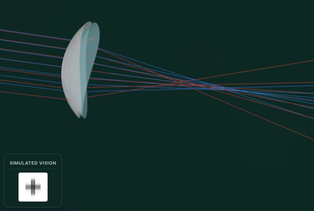
The final model combines everything above: the six layers, a toric shape with its two curvatures shown in clinical diopters (K=337.5/R in mm), the aspheric Q flattening, and adjustable thickness. The light rays are a simplified trace through the front surface only. They show how astigmatism and asphericity smear a focus, without pretending to be a full optical simulation.
import React, { useEffect, useRef, useState } from "react";
import * as THREE from "three";
import { OrbitControls } from "three/examples/jsm/controls/OrbitControls.js";
import { ErrorBoundary } from "../ErrorBoundary";
import { observeThreeResize } from "../threeResize";

function CorneaMasterInner() {
	const mountRef = useRef<HTMLDivElement>(null);

	// State for interactive sliders
	const [k1, setK1] = useState(7.8);
	const [k2, setK2] = useState(7.8);
	const [asphericity, setAsphericity] = useState(-0.26);
	const [thickness, setThickness] = useState(0.55);

	const sceneRef = useRef<THREE.Scene | null>(null);
	const geoRef = useRef<{
		mesh: THREE.Mesh;
		postMesh: THREE.Mesh;
		wire: THREE.Mesh;
		postWire: THREE.Mesh;
		rays: THREE.Group;
	} | null>(null);

	useEffect(() => {
		if (!mountRef.current) return;

		const scene = new THREE.Scene();
		sceneRef.current = scene;
		const camera = new THREE.PerspectiveCamera(45, 1, 0.1, 100);
		// Move camera to view the entire ray tracing from left (-X) to right (+X)
		camera.position.set(5.374, 18.032, 29.638);
		camera.zoom = 1;

		const ambientLight = new THREE.AmbientLight(0xffffff, 0.6);
		scene.add(ambientLight);
		const dirLight = new THREE.DirectionalLight(0xffffff, 1.0);
		dirLight.position.set(10, 15, 20);
		scene.add(dirLight);

		const renderer = new THREE.WebGLRenderer({ antialias: true, alpha: true });
		renderer.setPixelRatio(window.devicePixelRatio);
		renderer.setClearColor(0x000000, 0);
		mountRef.current.appendChild(renderer.domElement);

		const orbit = new OrbitControls(camera, renderer.domElement);
		orbit.enableDamping = true;
		orbit.dampingFactor = 0.05;
		orbit.target.set(10, 0, 0);

		// Handle responsive resize
		const resizeObserver = observeThreeResize(
			mountRef.current,
			renderer,
			camera,
		);

		const group = new THREE.Group();
		scene.add(group);

		// Provide a dummy geometry with valid attributes so WireframeGeometry doesn't crash on mount
		const geometry = new THREE.SphereGeometry(5, 32, 32);
		// Create Anterior Mesh
		const meshMat = new THREE.MeshPhysicalMaterial({
			color: 0xffffff,
			transmission: 0.9,
			opacity: 1,
			metalness: 0,
			roughness: 0,
			ior: 1.376,
			thickness: 1.0,
			side: THREE.DoubleSide,
			transparent: true,
		});
		const mesh = new THREE.Mesh(geometry, meshMat);
		mesh.rotation.y = Math.PI / 2; // Apex points left (-X)
		group.add(mesh);

		// Create Posterior Mesh (to show CCT)
		const postMat = new THREE.MeshPhysicalMaterial({
			color: 0xe0f7fa, // slight blue tint
			transmission: 0.95,
			opacity: 0.8,
			metalness: 0,
			roughness: 0.1,
			ior: 1.336, // aqueous humor
			side: THREE.DoubleSide,
			transparent: true,
		});
		const postMesh = new THREE.Mesh(geometry, postMat);
		postMesh.rotation.y = Math.PI / 2; // Apex points left (-X)
		postMesh.position.x = thickness; // Shift inwards (towards +X) by CCT
		group.add(postMesh);

		// Wireframes
		const wireMat = new THREE.LineBasicMaterial({
			color: 0xffffff,
			transparent: true,
			opacity: 0.1,
		});
		const wireframe = new THREE.LineSegments(
			new THREE.WireframeGeometry(geometry),
			wireMat,
		);
		mesh.add(wireframe);

		const postWireMat = new THREE.LineBasicMaterial({
			color: 0x00bcd4,
			transparent: true,
			opacity: 0.1,
		});
		const postWireframe = new THREE.LineSegments(
			new THREE.WireframeGeometry(geometry),
			postWireMat,
		);
		postMesh.add(postWireframe);

		const rays = new THREE.Group();
		group.add(rays);

		geoRef.current = {
			mesh,
			postMesh,
			wire: wireframe,
			postWire: postWireframe,
			rays,
		};

		let animationFrameId: number;
		const animate = () => {
			animationFrameId = requestAnimationFrame(animate);
			orbit.update();
			renderer.render(scene, camera);

			const debugEl = mountRef.current
				?.closest(".interactive-viewer")
				?.querySelector(".debug-output");
			if (debugEl) {
				const state = {
					camera: {
						position: {
							x: Number(camera.position.x.toFixed(3)),
							y: Number(camera.position.y.toFixed(3)),
							z: Number(camera.position.z.toFixed(3)),
						},
						rotation: {
							x: Number(camera.rotation.x.toFixed(3)),
							y: Number(camera.rotation.y.toFixed(3)),
							z: Number(camera.rotation.z.toFixed(3)),
						},
						zoom: Number(camera.zoom.toFixed(3)),
					},
					target: {
						x: Number(orbit.target.x.toFixed(3)),
						y: Number(orbit.target.y.toFixed(3)),
						z: Number(orbit.target.z.toFixed(3)),
					},
				};
				(debugEl as HTMLElement).innerText = JSON.stringify(state, null, 2);
			}
		};
		animate();

		return () => {
			cancelAnimationFrame(animationFrameId);
			resizeObserver.disconnect();
			if (mountRef.current && renderer.domElement.parentNode) {
				mountRef.current.removeChild(renderer.domElement);
			}
			renderer.dispose();
			geometry.dispose();
			meshMat.dispose();
			postMat.dispose();
			wireMat.dispose();
			postWireMat.dispose();
		};
	}, []);

	const [sidebarOpen, setSidebarOpen] = useState(true);

	// Update geometry when sliders change
	useEffect(() => {
		if (!geoRef.current) return;

		const { mesh, postMesh, rays } = geoRef.current;

		const scale = 1.0;
		const Rx = k1 * scale;
		const Ry = k2 * scale;
		const cx = 1 / Rx;
		const cy = 1 / Ry;
		const k = asphericity;
		const maxR = 5.0;

		const radialSegments = 64;
		const angularSegments = 64;

		const geometry = new THREE.BufferGeometry();
		const vertices = [];
		const indices = [];

		for (let i = 0; i <= radialSegments; i++) {
			const r = (i / radialSegments) * maxR;

			for (let j = 0; j <= angularSegments; j++) {
				const theta = (j / angularSegments) * Math.PI * 2;
				const x = r * Math.cos(theta);
				const y = r * Math.sin(theta);

				let z = 0;
				if (r > 0) {
					const denom =
						1 + Math.sqrt(1 - (1 + k) * (cx * cx * x * x + cy * cy * y * y));
					if (
						!isNaN(denom) &&
						denom !== 0 &&
						1 - (1 + k) * (cx * cx * x * x + cy * cy * y * y) >= 0
					) {
						z = (cx * x * x + cy * y * y) / denom;
					} else {
						z = vertices[vertices.length - 3] || 0;
					}
				}

				vertices.push(x, y, z);
			}
		}

		for (let i = 0; i < radialSegments; i++) {
			for (let j = 0; j < angularSegments; j++) {
				const a = i * (angularSegments + 1) + j;
				const b = a + 1;
				const cIdx = (i + 1) * (angularSegments + 1) + j;
				const d = cIdx + 1;
				indices.push(a, b, d);
				indices.push(a, d, cIdx);
			}
		}

		geometry.setAttribute(
			"position",
			new THREE.Float32BufferAttribute(vertices, 3),
		);
		geometry.setIndex(indices);
		geometry.computeVertexNormals();

		const oldGeo = mesh.geometry;
		mesh.geometry = geometry;
		postMesh.geometry = geometry;

		// Update wireframes
		mesh.children[0].geometry = new THREE.WireframeGeometry(geometry);
		postMesh.children[0].geometry = new THREE.WireframeGeometry(geometry);

		// Update CCT offset
		postMesh.position.x = thickness;

		oldGeo.dispose();

		while (rays.children.length > 0) {
			rays.remove(rays.children[0]);
		}

		// Draw Major and Minor Axes on the Cornea
		// Local X-axis meridian (World Z after Math.PI/2 rotation)
		const ptsX = [];
		for (let r = -maxR; r <= maxR; r += 0.1) {
			const denom = 1 + Math.sqrt(1 - (1 + k) * cx * cx * r * r);
			const z = denom > 0 && !isNaN(denom) ? (cx * r * r) / denom : 0;
			ptsX.push(new THREE.Vector3(z, 0, -r)); // World coordinates
		}
		const lineXGeo = new THREE.BufferGeometry().setFromPoints(ptsX);
		const lineXMat = new THREE.LineBasicMaterial({
			color: 0x3b82f6,
			depthTest: false,
			linewidth: 3,
		}); // Blue
		rays.add(new THREE.Line(lineXGeo, lineXMat));

		// Local Y-axis meridian (World Y after Math.PI/2 rotation)
		const ptsY = [];
		for (let r = -maxR; r <= maxR; r += 0.1) {
			const denom = 1 + Math.sqrt(1 - (1 + k) * cy * cy * r * r);
			const z = denom > 0 && !isNaN(denom) ? (cy * r * r) / denom : 0;
			ptsY.push(new THREE.Vector3(z, r, 0)); // World coordinates
		}
		const lineYGeo = new THREE.BufferGeometry().setFromPoints(ptsY);
		const lineYMat = new THREE.LineBasicMaterial({
			color: 0xef4444,
			depthTest: false,
			linewidth: 3,
		}); // Red
		rays.add(new THREE.Line(lineYGeo, lineYMat));

		// Ray tracing - Sturm's Conoid (Two orthogonal planes)
		const numRays = 7;
		const spread = 8.0;

		// 1. Vertical fan (Red rays, Y-axis)
		for (let i = 0; i < numRays; i++) {
			const offset = -spread / 2 + (spread / (numRays - 1)) * i;
			const pts = [];
			pts.push(new THREE.Vector3(-15, offset, 0));

			let hitZ = 0;
			const rootTerm = 1 - (1 + k) * cy * cy * offset * offset;
			if (rootTerm >= 0) {
				hitZ = (cy * offset * offset) / (1 + Math.sqrt(rootTerm));
			} else {
				pts.push(new THREE.Vector3(35, offset, 0));
				const lineMat = new THREE.LineBasicMaterial({
					color: 0xef4444,
					opacity: 0.1,
					transparent: true,
				});
				rays.add(
					new THREE.Line(
						new THREE.BufferGeometry().setFromPoints(pts),
						lineMat,
					),
				);
				continue;
			}

			pts.push(new THREE.Vector3(hitZ, offset, 0));
			const f0 = k2 / 0.376;
			const aberration = (k + 1) * 0.25 * (offset * offset);
			const focusX = f0 - aberration;
			pts.push(new THREE.Vector3(focusX, 0, 0));

			const slope = (0 - offset) / (focusX - hitZ);
			const endX = 35;
			const endY = slope * (endX - focusX);
			pts.push(new THREE.Vector3(endX, endY, 0));

			const lineMat = new THREE.LineBasicMaterial({
				color: 0xef4444,
				opacity: 0.6,
				transparent: true,
			});
			rays.add(
				new THREE.Line(new THREE.BufferGeometry().setFromPoints(pts), lineMat),
			);
		}

		// 2. Horizontal fan (Blue rays, Z-axis)
		for (let i = 0; i < numRays; i++) {
			const offset = -spread / 2 + (spread / (numRays - 1)) * i;
			const pts = [];
			pts.push(new THREE.Vector3(-15, 0, offset));

			let hitZ = 0;
			const rootTerm = 1 - (1 + k) * cx * cx * offset * offset;
			if (rootTerm >= 0) {
				hitZ = (cx * offset * offset) / (1 + Math.sqrt(rootTerm));
			} else {
				pts.push(new THREE.Vector3(35, 0, offset));
				const lineMat = new THREE.LineBasicMaterial({
					color: 0x3b82f6,
					opacity: 0.1,
					transparent: true,
				});
				rays.add(
					new THREE.Line(
						new THREE.BufferGeometry().setFromPoints(pts),
						lineMat,
					),
				);
				continue;
			}

			pts.push(new THREE.Vector3(hitZ, 0, offset));
			const f0 = k1 / 0.376;
			const aberration = (k + 1) * 0.25 * (offset * offset);
			const focusX = f0 - aberration;
			pts.push(new THREE.Vector3(focusX, 0, 0));

			const slope = (0 - offset) / (focusX - hitZ);
			const endX = 35;
			const endZ = slope * (endX - focusX);
			pts.push(new THREE.Vector3(endX, 0, endZ));

			const lineMat = new THREE.LineBasicMaterial({
				color: 0x3b82f6,
				opacity: 0.6,
				transparent: true,
			});
			rays.add(
				new THREE.Line(new THREE.BufferGeometry().setFromPoints(pts), lineMat),
			);
		}
	}, [k1, k2, asphericity, thickness]);

	// Calculate directional blur for simulated vision
	const idealK = 7.8;
	const idealQ = -0.26;
	const idealThickness = 0.55; // 550 microns is normal CCT

	const errorK1 = Math.abs(k1 - idealK);
	const errorK2 = Math.abs(k2 - idealK);
	const aberrationError = Math.abs(asphericity - idealQ);
	const thicknessError = Math.abs(thickness - idealThickness);

	// Astigmatism causes directional blur (K1 = X-axis, K2 = Y-axis).
	// Aberration causes a soft glow.
	// Thickness deviations cause a slight myopic/hyperopic spherical shift (uniform blur).
	// Multiplied by 60 to make the microscopic effect visually apparent in the UI.
	const blurX = errorK1 * 2.5 + aberrationError * 5.0 + thicknessError * 60.0;
	const blurY = errorK2 * 2.5 + aberrationError * 5.0 + thicknessError * 60.0;

	const renderCrossOptotype = (filterId: string) => (
		<svg
			width="40"
			height="40"
			viewBox="0 0 100 100"
			style={{ filter: `url(#${filterId})` }}
		>
			{/* Vertical arm */}
			<line
				x1="42"
				y1="10"
				x2="42"
				y2="90"
				stroke="currentColor"
				strokeWidth="3"
				strokeLinecap="round"
			/>
			<line
				x1="50"
				y1="10"
				x2="50"
				y2="90"
				stroke="currentColor"
				strokeWidth="3"
				strokeLinecap="round"
			/>
			<line
				x1="58"
				y1="10"
				x2="58"
				y2="90"
				stroke="currentColor"
				strokeWidth="3"
				strokeLinecap="round"
			/>
			{/* Horizontal arm */}
			<line
				x1="10"
				y1="42"
				x2="90"
				y2="42"
				stroke="currentColor"
				strokeWidth="3"
				strokeLinecap="round"
			/>
			<line
				x1="10"
				y1="50"
				x2="90"
				y2="50"
				stroke="currentColor"
				strokeWidth="3"
				strokeLinecap="round"
			/>
			<line
				x1="10"
				y1="58"
				x2="90"
				y2="58"
				stroke="currentColor"
				strokeWidth="3"
				strokeLinecap="round"
			/>
		</svg>
	);

	return (
		<div className="relative w-full h-[500px] md:h-[600px] bg-transparent overflow-hidden rounded-lg group">
			<div
				ref={mountRef}
				className="absolute inset-0 cursor-grab active:cursor-grabbing"
			/>

			{/* Simulated Vision Overlay */}
			<div className="absolute bottom-4 left-4 z-20 flex flex-col items-center bg-card/80 backdrop-blur-md border border-border p-3 rounded-xl shadow-lg pointer-events-none">
				<span className="text-[10px] uppercase tracking-wider font-bold text-muted-foreground border-b border-border/50 pb-1 w-full text-center mb-2">
					Simulated Vision
				</span>
				<div className="bg-white p-2 rounded flex items-center justify-center w-16 h-16 relative overflow-hidden">
					<svg width="0" height="0" className="absolute">
						<defs>
							<filter id="astigBlurCore">
								<feGaussianBlur
									stdDeviation={`${blurX * 0.3},${blurY * 0.3}`}
								/>
							</filter>
							<filter id="astigBlurHalo">
								<feGaussianBlur stdDeviation={`${blurX},${blurY}`} />
							</filter>
						</defs>
					</svg>

					<div
						className="absolute inset-0 flex items-center justify-center text-black"
						style={{ opacity: 0.6 }}
					>
						{renderCrossOptotype("astigBlurHalo")}
					</div>
					<div className="absolute inset-0 flex items-center justify-center text-black">
						{renderCrossOptotype("astigBlurCore")}
					</div>
				</div>
			</div>

			<button
				onClick={() => setSidebarOpen(!sidebarOpen)}
				className="absolute top-4 right-4 z-20 px-2 py-1.5 rounded-md bg-transparent hover:bg-background/40 backdrop-blur-sm border border-transparent hover:border-border/30 transition-all text-[10px] uppercase tracking-wider font-bold flex items-center gap-1.5 text-muted-foreground hover:text-foreground"
			>
				<svg
					xmlns="http://www.w3.org/2000/svg"
					width="12"
					height="12"
					viewBox="0 0 24 24"
					fill="none"
					stroke="currentColor"
					strokeWidth="2"
					strokeLinecap="round"
					strokeLinejoin="round"
				>
					<line x1="4" y1="21" x2="4" y2="14"></line>
					<line x1="4" y1="10" x2="4" y2="3"></line>
					<line x1="12" y1="21" x2="12" y2="12"></line>
					<line x1="12" y1="8" x2="12" y2="3"></line>
					<line x1="20" y1="21" x2="20" y2="16"></line>
					<line x1="20" y1="12" x2="20" y2="3"></line>
					<line x1="1" y1="14" x2="7" y2="14"></line>
					<line x1="9" y1="8" x2="15" y2="8"></line>
					<line x1="17" y1="16" x2="23" y2="16"></line>
				</svg>
				{sidebarOpen ? "Hide" : "Controls"}
			</button>

			{/* Control Panel */}
			<div
				className={`absolute top-12 right-4 w-[220px] p-3 border border-border/10 hover:border-border/30 bg-background/30 hover:bg-background/80 backdrop-blur-md rounded-xl shadow-sm hover:shadow-lg flex flex-col gap-3 text-xs z-10 transition-all duration-300 origin-top-right ${sidebarOpen ? "scale-100 opacity-100 pointer-events-auto" : "scale-95 opacity-0 pointer-events-none"}`}
			>
				<div>
					<label className="flex justify-between mb-1 font-medium text-foreground/80">
						<span>K1 (Flat)</span>
						<span className="text-muted-foreground">{k1.toFixed(1)} mm</span>
					</label>
					<input
						type="range"
						min="6.0"
						max="9.0"
						step="0.1"
						value={k1}
						onChange={(e) => setK1(parseFloat(e.target.value))}
						className="w-full h-1 bg-muted rounded-lg appearance-none cursor-pointer accent-primary"
					/>
				</div>
				<div>
					<label className="flex justify-between mb-1 font-medium text-foreground/80">
						<span>K2 (Steep)</span>
						<span className="text-muted-foreground">{k2.toFixed(1)} mm</span>
					</label>
					<input
						type="range"
						min="6.0"
						max="9.0"
						step="0.1"
						value={k2}
						onChange={(e) => setK2(parseFloat(e.target.value))}
						className="w-full h-1 bg-muted rounded-lg appearance-none cursor-pointer accent-primary"
					/>
				</div>
				<div>
					<label className="flex justify-between mb-1 font-medium text-foreground/80">
						<span>Asphericity (Q)</span>
						<span className="text-muted-foreground">
							{asphericity.toFixed(2)}
						</span>
					</label>
					<input
						type="range"
						min="-1.0"
						max="1.0"
						step="0.01"
						value={asphericity}
						onChange={(e) => setAsphericity(parseFloat(e.target.value))}
						className="w-full h-1 bg-muted rounded-lg appearance-none cursor-pointer accent-primary"
					/>
				</div>
				<div>
					<label className="flex justify-between mb-1 font-medium text-foreground/80">
						<span>Thickness</span>
						<span className="text-muted-foreground">
							{Math.round(thickness * 1000)} µm
						</span>
					</label>
					<input
						type="range"
						min="0.4"
						max="0.7"
						step="0.01"
						value={thickness}
						onChange={(e) => setThickness(parseFloat(e.target.value))}
						className="w-full h-1 bg-muted rounded-lg appearance-none cursor-pointer accent-primary"
					/>
				</div>
			</div>
		</div>
	);
}

export default function CorneaMaster() {
	return (
		<ErrorBoundary>
			<CorneaMasterInner />
		</ErrorBoundary>
	);
}A hyperrealistic finish
The models above are built to explain, so they favor clear colors and diagrams over looking real. To close the loop, here is just the cornea dressed for realism: the same aspheric shape, now a clear meniscus dome (an aspheric front surface with a steeper back surface) made of a glassy transmission material with a clearcoat, refracting a synthetic studio environment. The bright highlight riding on the surface is the corneal reflection an optician calls Purkinje image P1, the same reflection Placido rings rely on. Drag to move it around the dome.
import React, { useEffect, useRef } from "react";
import * as THREE from "three";
import { OrbitControls } from "three/examples/jsm/controls/OrbitControls.js";
import { RoomEnvironment } from "three/examples/jsm/environments/RoomEnvironment.js";
import { RectAreaLightUniformsLib } from "three/examples/jsm/lights/RectAreaLightUniformsLib.js";
import { observeThreeResize } from "../threeResize";

// Average anatomical values (mm). Built at true scale, framed by the camera.
const R_ANT = 7.8; // anterior radius of curvature
const Q_ANT = -0.26; // anterior asphericity (prolate)
const R_POST = 6.5; // posterior radius (steeper)
const Q_POST = -0.4;
const CCT = 0.55; // central corneal thickness
const SEMI = 5.0; // optical-zone semi-diameter

function sag(r: number, R: number, Q: number) {
	const c = 1 / R;
	const root = 1 - (1 + Q) * c * c * r * r;
	return root >= 0 ? (c * r * r) / (1 + Math.sqrt(root)) : c * r * r;
}

// Meniscus cornea: aspheric front, steeper back, joined at the rim, lathed into
// a closed glassy solid.
function corneaLathe() {
	const steps = 128;
	const pts: THREE.Vector2[] = [];
	for (let i = 0; i <= steps; i++) {
		const r = (i / steps) * SEMI;
		pts.push(new THREE.Vector2(r, -sag(r, R_ANT, Q_ANT)));
	}
	for (let i = steps; i >= 0; i--) {
		const r = (i / steps) * SEMI;
		pts.push(new THREE.Vector2(r, -CCT - sag(r, R_POST, Q_POST)));
	}
	const geo = new THREE.LatheGeometry(pts, 160);
	geo.center();
	geo.computeVertexNormals();
	return geo;
}

// Surrounding studio world: dark gradient + faint grid on the inside of a big
// sphere, visible from any angle and something for the cornea to refract.
function worldTexture() {
	const w = 2048,
		h = 1024;
	const c = document.createElement("canvas");
	c.width = w;
	c.height = h;
	const ctx = c.getContext("2d")!;
	const g = ctx.createLinearGradient(0, 0, 0, h);
	g.addColorStop(0, "#0a1512");
	g.addColorStop(0.5, "#16241f");
	g.addColorStop(1, "#05080a");
	ctx.fillStyle = g;
	ctx.fillRect(0, 0, w, h);
	ctx.strokeStyle = "rgba(130,180,190,0.15)";
	ctx.lineWidth = 2;
	for (let i = 0; i <= 48; i++) {
		ctx.beginPath();
		ctx.moveTo((i / 48) * w, 0);
		ctx.lineTo((i / 48) * w, h);
		ctx.stroke();
	}
	for (let j = 0; j <= 24; j++) {
		ctx.beginPath();
		ctx.moveTo(0, (j / 24) * h);
		ctx.lineTo(w, (j / 24) * h);
		ctx.stroke();
	}
	const tex = new THREE.CanvasTexture(c);
	tex.colorSpace = THREE.SRGBColorSpace;
	return tex;
}

// Smooth grayscale noise varying the tear-film thickness (oil-slick bands).
function thicknessMap() {
	const small = document.createElement("canvas");
	small.width = small.height = 16;
	const sctx = small.getContext("2d")!;
	const img = sctx.createImageData(16, 16);
	for (let i = 0; i < img.data.length; i += 4) {
		const v = Math.floor(Math.random() * 255);
		img.data[i] = img.data[i + 1] = img.data[i + 2] = v;
		img.data[i + 3] = 255;
	}
	sctx.putImageData(img, 0, 0);
	const big = document.createElement("canvas");
	big.width = big.height = 256;
	const bctx = big.getContext("2d")!;
	bctx.imageSmoothingEnabled = true;
	bctx.drawImage(small, 0, 0, 256, 256);
	return new THREE.CanvasTexture(big);
}

export default function CorneaRealistic() {
	const mountRef = useRef<HTMLDivElement>(null);

	useEffect(() => {
		if (!mountRef.current) return;

		const scene = new THREE.Scene();
		const camera = new THREE.PerspectiveCamera(35, 1, 0.1, 200);
		camera.position.set(6, 4, 18);

		const renderer = new THREE.WebGLRenderer({ antialias: true, alpha: true });
		renderer.setPixelRatio(Math.min(window.devicePixelRatio, 2));
		renderer.setClearColor(0x000000, 0);
		renderer.toneMapping = THREE.ACESFilmicToneMapping;
		renderer.toneMappingExposure = 1.15;
		mountRef.current.appendChild(renderer.domElement);

		const pmrem = new THREE.PMREMGenerator(renderer);
		const envTex = pmrem.fromScene(new RoomEnvironment(), 0.04).texture;
		scene.environment = envTex;

		const controls = new OrbitControls(camera, renderer.domElement);
		controls.enableDamping = true;
		controls.dampingFactor = 0.05;
		controls.enablePan = false;
		controls.minDistance = 12;
		controls.maxDistance = 45;

		const resizeObserver = observeThreeResize(
			mountRef.current,
			renderer,
			camera,
		);

		const dir = new THREE.DirectionalLight(0xffffff, 2.4);
		dir.position.set(-6, 9, 12);
		scene.add(dir);
		scene.add(new THREE.AmbientLight(0xffffff, 0.25));

		// Softbox-style light: reflects off the wet surface as a crisp catchlight.
		RectAreaLightUniformsLib.init();
		const softbox = new THREE.RectAreaLight(0xffffff, 6, 7, 4);
		softbox.position.set(-7, 8, 11);
		softbox.lookAt(0, 0, 0);
		scene.add(softbox);

		// Surrounding world.
		const worldTex = worldTexture();
		const world = new THREE.Mesh(
			new THREE.SphereGeometry(60, 48, 48),
			new THREE.MeshBasicMaterial({ map: worldTex, side: THREE.BackSide }),
		);
		scene.add(world);

		const geo = corneaLathe();
		const filmMap = thicknessMap();
		filmMap.wrapS = filmMap.wrapT = THREE.RepeatWrapping;
		const mat = new THREE.MeshPhysicalMaterial({
			color: 0xffffff,
			transmission: 1.0,
			thickness: CCT,
			ior: 1.376,
			// Chromatic dispersion: rainbow fringing at refracted edges.
			dispersion: 0.4,
			roughness: 0.015,
			metalness: 0.0,
			// Wet surface reflection.
			clearcoat: 1.0,
			clearcoatRoughness: 0.015,
			specularIntensity: 1.0,
			envMapIntensity: 1.3,
			// Tear-film thin-film interference (oil-slick sheen).
			iridescence: 1.0,
			iridescenceIOR: 1.32,
			iridescenceThicknessRange: [80, 420],
			iridescenceThicknessMap: filmMap,
			// A touch of anisotropy from the corneal collagen grain.
			anisotropy: 0.2,
			transparent: true,
			attenuationColor: new THREE.Color(0xd6ecff),
			attenuationDistance: 12,
			side: THREE.DoubleSide,
		});
		const cornea = new THREE.Mesh(geo, mat);
		cornea.rotation.x = -Math.PI / 2; // axis toward the camera (+Z)
		scene.add(cornea);

		let animationFrameId: number;
		const animate = () => {
			animationFrameId = requestAnimationFrame(animate);

			// Slowly drift the tear-film thickness so the sheen moves like tears
			// spreading across the surface after a blink.
			filmMap.offset.x += 0.0006;
			filmMap.offset.y += 0.0003;

			controls.update();
			renderer.render(scene, camera);

			const debugEl = mountRef.current
				?.closest(".interactive-viewer")
				?.querySelector(".debug-output");
			if (debugEl) {
				const state = {
					camera: {
						position: {
							x: +camera.position.x.toFixed(3),
							y: +camera.position.y.toFixed(3),
							z: +camera.position.z.toFixed(3),
						},
					},
					target: {
						x: +controls.target.x.toFixed(3),
						y: +controls.target.y.toFixed(3),
						z: +controls.target.z.toFixed(3),
					},
				};
				(debugEl as HTMLElement).innerText = JSON.stringify(state, null, 2);
			}
		};
		animate();

		return () => {
			cancelAnimationFrame(animationFrameId);
			resizeObserver.disconnect();
			if (mountRef.current && renderer.domElement.parentNode) {
				mountRef.current.removeChild(renderer.domElement);
			}
			geo.dispose();
			mat.dispose();
			filmMap.dispose();
			worldTex.dispose();
			world.geometry.dispose();
			(world.material as THREE.Material).dispose();
			envTex.dispose();
			pmrem.dispose();
			renderer.dispose();
		};
	}, []);

	return (
		<div className="relative w-full h-[500px] md:h-[600px] bg-transparent overflow-hidden rounded-lg group">
			<div className="absolute top-4 left-4 z-10 flex flex-col items-start gap-1 pointer-events-none bg-card/80 backdrop-blur-md border border-border p-3 rounded-xl shadow-lg">
				<span className="text-[10px] uppercase tracking-wider font-bold text-muted-foreground mb-1 border-b border-border/50 pb-1 w-full text-center">
					Hyperrealistic Cornea
				</span>
				<div className="text-[10px] text-muted-foreground max-w-[220px] leading-relaxed">
					Built to average anatomy: R<sub>ant</sub> 7.8 mm, R<sub>post</sub> 6.5
					mm, Q −0.26, thickness 0.55 mm. Glassy transmission with dispersion, a
					drifting tear-film sheen, and reflections. Drag to orbit.
				</div>
			</div>
			<div
				ref={mountRef}
				className="w-full h-full cursor-grab active:cursor-grabbing"
			/>
		</div>
	);
}That gives us a solid front surface for the eye.
References
- Young, T. (1801). On the Mechanism of the Eye. Philosophical Transactions of the Royal Society of London, 91, 23-88.
- Helmholtz, H. von (1854). Ueber die Accommodation des Auges. Graefes Archiv für Ophthalmologie, 1(2), 1-74.
- Atchison, D. A., & Smith, G. (2000). Optics of the Human Eye. Butterworth-Heinemann, Oxford, Chapter 2.
- Maurice, D. M. (1957). The structure and transparency of the cornea. Journal of Physiology, 136(2), 263-286.
- Benedek, G. B. (1971). Theory of transparency of the eye. Applied Optics, 10(3), 459-473.
- Dua, H. S., Faraj, L. A., Said, D. G., Gray, T., & Lowe, J. (2013). Human Corneal Anatomy Redefined: A Novel Layer (Dua’s Layer). Ophthalmology, 120(9), 1778-1785.
- Plácido, A. (1880). Novo instrumento para analyse da curvatura da cornea. Periodico Ophthalmologico, 2(5), 44-49, Lisbon.